
AUGUST 19, 2026  — Mayo Clinic researchers have developed and externally validated an artificial intelligence (AI) model that can identify a potentially significant heart obstruction from routine ultrasound videos without relying on specialized Doppler imaging. The technology could help clinicians flag patients with hypertrophic cardiomyopathy (HCM) who may need additional testing, particularly in settings where specialized echocardiography expertise is limited. Study findings are published in Circulation: Cardiovascular Imaging.

HCM is a genetic condition that causes the heart muscle to become abnormally thick. About two-thirds of these patients develop left ventricular outflow tract (LVOT) obstruction, which restricts blood leaving the heart, causing symptoms such as chest pain and shortness of breath with exertion or when lying flat. Knowing which patients develop LVOT obstruction is important because it influences treatment decisions and long-term management for patients with HCM.

“Measuring LVOT obstruction typically requires Doppler echocardiography, which depends on precise ultrasound-beam alignment and operator expertise,” says Imon Banerjee, Ph.D., an AI researcher at Mayo Clinic in Phoenix and senior author of the study. “We wanted to determine whether AI could recognize subtle patterns that are imperceptible to the human eye in routinely acquired B-mode ultrasound videos and identify patients with LVOT obstruction earlier, enabling timely confirmatory Doppler evaluation and referral when appropriate.”

The study included 1,833 patients in the Mayo Clinic cohort. The model was tested in 275 patients and externally validated in 46 patients from a hospital in South Korea. The AI model used only resting, non-Doppler ultrasound videos to predict whether a patient had a potentially significant obstruction to blood leaving the heart. Researchers found that combining information from three standard ultrasound views improved the model’s ability to distinguish patients with elevated LVOT gradients. The model also helped identify obstruction that may only appear when the heart is under stress.

The model maintained strong performance in the South Korean group despite substantial differences between that population and the patients used to develop the model, supporting further study of the technology across different patient populations and clinical settings.

In a subset of cases, the AI model identified obstruction more accurately than two expert echocardiographers who reviewed the same non-Doppler images. The findings highlight how difficult it can be to recognize LVOT obstruction from routine two-dimensional images without Doppler measurements.

“This technology is intended to complement, not replace, Doppler echocardiography,” Dr. Banerjee says. “By enabling earlier identification of patients with potential LVOT obstruction, it could escalate timely detection and prompt confirmatory Doppler measurements, stress testing, or referral to an HCM specialty center. It also could support evaluation using portable ultrasound or in settings where comprehensive Doppler assessment may not be readily available, helping expand access to earlier screening and risk assessment.”

Dr. Banerjee notes that the next steps include additional prospective validation across broader clinical settings, ultrasound platforms and patient populations.

The list of authors and disclosures may be found in the article, Beyond Doppler: Scalable AI Detection of LVOT Obstruction in HCM. This study received no external funding.

About Mayo Clinic

Mayo Clinic is a nonprofit organization committed to innovation in clinical practice, education and research, and providing compassion, expertise and answers to everyone who needs healing. Visit the Mayo Clinic News Network for additional Mayo Clinic news.


BY: Brittany Cordeiro

AUGUST 20, 2026  — Mayo Clinic researchers have estimated the risk that pancreatic cysts with worrisome or high-risk features will progress to pancreatic cancer or advanced precancerous changes within three years. The findings, published in Gastroenterology, could help physicians and patients better assess risk and make more informed decisions about whether to monitor a cyst or consider surgery.

The prospective study found that risk increased with the number of worrisome features, or findings that the cyst needs closer monitoring. Patients whose cysts had one worrisome feature had about a 2% chance of developing pancreatic cancer or advanced precancerous changes within three years. The risk rose to 4% for those with two worrisome features and 18% for those with three or more.

“This information will help us counsel patients. If we want to intercept pancreatic cancer while also sparing patients from unnecessary interventions, we must really know what is likely to become cancer and what is not,” says Shounak Majumder, M.D., lead author and a gastroenterologist at Mayo Clinic.

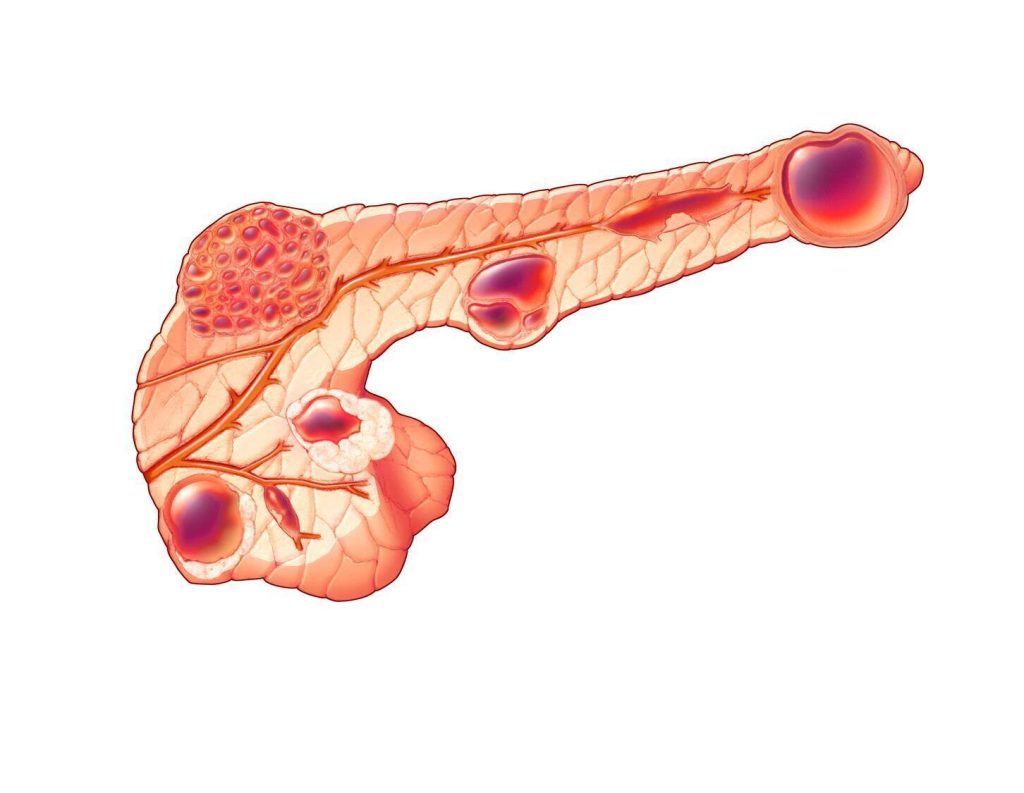
Mucinous cysts in the pancreas

What does pancreatic cyst risk mean for patients? 

Pancreatic cysts are common and often discovered during imaging for an unrelated reason. Most do not become cancerous, but certain features seen on imaging can indicate a greater risk. Pancreatic cysts are classified as having high-risk features, worrisome features or neither.

Pancreatic cysts without worrisome or high-risk features have a relatively low likelihood of progression to cancer. Cysts with high-risk features may prompt physicians to recommend surgery because of greater concern about cancer or advanced precancerous changes. Cysts with worrisome features often are monitored closely, although some may also require surgery.

“Sometimes, we are unable to reliably determine whether a cyst will become pancreatic cancer or not,” Dr. Majumder says. “That uncertainty creates a lot of anxiety for some patients undergoing surveillance, and some will opt for surgery.”

More precise estimates of progression risk can help patients and physicians weigh the potential benefits of surveillance and surgery.

.

How did researchers determine pancreatic cancer risk? 

The study followed 230 people with pancreatic cysts that had worrisome or high-risk features. Unlike studies that look back at existing medical records or primarily include patients who underwent surgery, researchers enrolled participants and followed them over time, regardless of whether they chose surgery or surveillance.

Of the 230 participants, 32 underwent surgery within the first three months after enrollment, and 198 entered clinical surveillance. Among those monitored, 185 had cysts with worrisome features, and 13 had cysts with high-risk features.

The study found that patients with multiple worrisome features may represent a substantially higher-risk subgroup, while risk was not markedly elevated in patients with a single worrisome feature.

Pancreatic cancers diagnosed among patients under careful surveillance were detected at a very early stage, suggesting that surveillance of appropriately risk-stratified patients may provide an opportunity to catch pancreatic cancer early. The finding suggests that regular surveillance may be an appropriate approach for some patients rather than immediate surgery.

The three-year risk estimates also provide a benchmark for evaluating blood tests and designing studies of other approaches to early pancreatic cancer detection.

Can blood tests and biomarkers predict pancreatic cancer risk?

In a separate study led by Dr. Majumder and colleagues published in Clinical Gastroenterology and Hepatology, researchers examined CA 19-9, a blood biomarker that may be elevated in people with pancreatic cancer and is sometimes used to monitor pancreatic cysts.

An elevated CA 19-9 result alone did not reliably predict which patients with pancreatic cysts would develop cancer. Among patients with low-risk cysts who had an elevated CA 19-9 level, none developed pancreatic cancer during the study’s follow-up period.

The findings suggest that a blood biomarker test should be considered with other information about a patient’s health and cancer risk. They also underscore the need for new biomarker tests that can reliably detect the earliest stages of cancer in patients with pancreatic cysts.

The data from these studies could help researchers design larger studies and clinical trials of new approaches to the early detection and prevention of pancreatic cancer as part of a broader effort at Mayo Clinic known as the Precure Research initiative.

“The goal is more precise, personalized surveillance and an early detection approach that identifies patients who may benefit from earlier intervention while helping those at lower risk avoid unnecessary procedures and major surgery,” says Dr. Majumder.

For a complete list of authors, disclosures and funding, review the study.

###

About Mayo Clinic
Mayo Clinic is a nonprofit organization committed to innovation in clinical practice, education and research, and providing compassion, expertise and answers to everyone who needs healing. Visit the Mayo Clinic News Network for additional Mayo Clinic news.



AUGUST 21, 2026 – Imagine trying to read the fine print on a page through a fogged window. Important details are there, but they’re difficult to see clearly. Now imagine that window suddenly becoming crystal clear.

That’s the difference Memorial Healthcare System’s newest cardiac imaging technology can make for radiologists.

Memorial is the first healthcare system in South Florida to dedicate the world’s most advanced photon-counting CT scanner exclusively to cardiac imaging. The Siemens NAEOTOM Alpha provides physicians with an extraordinarily detailed look inside the heart, revealing tiny coronary arteries, dangerous plaque, and other abnormalities with a level of clarity that was not previously possible with conventional CT technology.

For physicians, that could mean the difference between identifying heart disease before it becomes a heart attack and discovering it only after permanent damage has occurred in their patients.

“Every day, we care for patients who come to us with chest pain, shortness of breath, or risk factors for heart disease,” said Shane Strum, interim CEO of Memorial Healthcare System. “This technology allows our physicians to see more, know more and act sooner. It’s another example of Memorial’s commitment to bringing the most advanced care available to the communities we serve.”

Heart disease remains the leading cause of death in the United States, often developing silently over many years. As plaque gradually builds inside the coronary arteries, blood flow to the heart becomes restricted. Detecting those changes early can give patients the opportunity to make lifestyle changes, begin medications, or receive treatment before a life-threatening event occurs.

Traditional CT scanners have transformed heart imaging over the past two decades, but they still have limitations. Calcium deposits can appear larger than they really are, making arteries seem more blocked than they actually are. Small vessels can be difficult to evaluate, and metal stents may obscure important details.

The NAEOTOM Alpha changes that.

Its photon-counting technology captures each individual X-ray photon, producing remarkably sharp, high-resolution images that allow physicians to see the heart in unprecedented detail. Tiny coronary arteries become clearer. Plaque can be characterized more accurately. Even patients with heavily calcified arteries or prior stents, often among the most difficult to image, can benefit from greater diagnostic confidence. The technology also delivers these images while using less radiation than many conventional CT systems.

Memorial further enhances those images with HeartFlow FFRCT, an AI-enabled technology that transforms the scan into a personalized three-dimensional model of the patient’s coronary arteries. Together, the technologies help physicians not only see potential blockages more clearly but also better understand whether those blockages are limiting blood flow to the heart, leading to more informed treatment decisions and in many cases, helping patients avoid unnecessary invasive procedures.

“Think of it as the difference between watching an old standard-definition television and today’s ultra-high-definition displays,” said Vamsi Pavuluri, MD, medical director of cardiac imaging operations at Memorial Cardiac and Vascular Institute. “Now we’re seeing with a level of clarity that wasn’t previously possible, while also gaining valuable information about how blood is flowing through the coronary arteries. That combination allows us to make more precise decisions about each patient’s care and determine who may benefit from an invasive coronary intervention and who needs medical therapy optimized.”

For many patients, the technology may also reduce the need for cardiac catheterization to simply determine whether a blockage exists.

Beyond diagnosing coronary artery disease, the scanner supports patients with congenital heart disease, structural heart conditions, valve disease, and those preparing for minimally invasive cardiac procedures. It also improves the evaluation of bypass grafts, coronary stents and complex heart anatomy.

For patients, the benefits are simple: earlier answers, more precise diagnoses and greater peace of mind.

The addition of this technology reflects Memorial’s continued investment in providing patients with access to the most advanced cardiovascular care available, close to home.

The NAEOTOM Alpha is located at the newly redesigned home of Memorial Cardiac and Vascular Institute at Memorial Hospital West in Pembroke Pines.


August 19, 2026 – Rehmann began his healthcare career in direct patient care as a firefighter/paramedic and an emergency department nurse before advancing into hospital leadership. He previously served as director of Emergency Services at HCA Florida Largo Hospital and returns after gaining additional leadership experience at Research Medical Center, part of HCA Healthcare, in Kansas City, Missouri, and most recently as vice president of nursing operations for the HCA Healthcare West Florida Division.

“J.R. brings meaningful patient care and hospital leadership experience to this role,” said Dr. Sebastian Strom, chief executive officer of HCA Florida Largo Hospital. “We are pleased to welcome him back to HCA Florida Largo Hospital and look forward to the positive impact he will have on our colleagues, patients and community.”

In his new role, Rehmann will help lead hospital operations with a focus on supporting care teams, strengthening operational performance and keeping patients at the center of care.

“I am honored to return to HCA Florida Largo Hospital and serve alongside the physicians, nurses and colleagues who make this hospital such an important part of our community,” Rehmann said. “My career began in emergency services and at the bedside, and that perspective will continue to guide my focus on supporting care teams, improving operations and serving patients with purpose.”


The $17 million facility, next to the entrance of the On Top of the World community and the Canopy Oak Center, will serve communities along the SR 200 corridor and western Marion County.

August 21, 2026 – HCA Florida Healthcare marked an important milestone in the construction of HCA Florida ER, its newest freestanding emergency room in Marion County, with a beam-signing ceremony on Aug. 21.

Colleagues from HCA Florida West Marion Hospital, HCA Florida Ocala Hospital, our other nearby freestanding emergency rooms, and members of the hospitals’ Board of Trustees participated in the beam-signing along with representatives of the Gainesville-based construction firm CPPI, which is building the $17 million facility. Traditionally, a beam-signing is a ceremonial event marking the placement of the final or highest structural beam.

Located at 8231 SW State Road 200, next to the entrance of the On Top of the World community and the Canopy Oak Center, HCA Florida ER will serve communities along the SR 200 corridor and other areas of western Marion County. The 11,000 square-foot facility will open in spring 2027 and be accessible by golf cart as well as other vehicles.

HCA Florida ER will be a full-service, fully staffed emergency room, employing 45 full-time healthcare professionals, including emergency physicians, nurses and other clinicians ready to serve the community with the same capabilities as the emergency departments at HCA Florida Ocala Hospital and HCA Florida West Marion Hospital.

During the beam-signing event, HCA Florida West Marion Hospital CEO Isaiah Zirkle read tributes to our ER teams from patients who praised our colleagues’ compassion, skill and dedication to providing the best of care. “This is why we do what we do, and we’re going to be able to provide that here,” said Zirkle. “We’re super excited for this to be our sixth freestanding emergency room in the Ocala/Marion County community.”