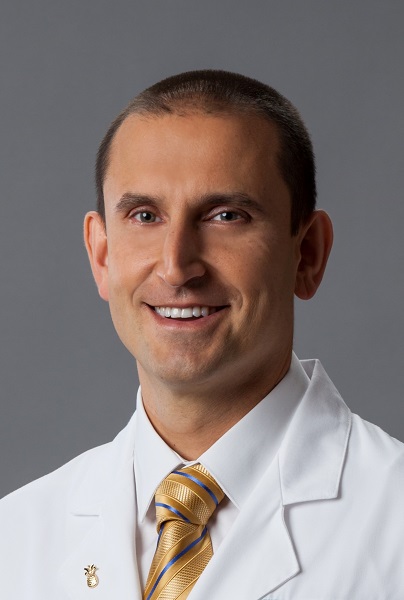


At Miami Neuroscience Institute, part of Baptist Health, neurosurgeons are taking a fresh look at how to reach deep brain tumors without the trauma often associated with traditional surgery. Led by Dr. Vitaly Siomin, director of cerebrovascular surgery and co-director of skull base surgery, the team’s recent study, published in Operative Neurosurgery, suggests that a simpler, less invasive approach could be both safe and effective.
Minimally invasive neurosurgery is a growing field that aims to access tumors and other lesions through smaller openings, specialized tools, and gentler tissue handling. The goal is to achieve the same precision and thoroughness as open surgery while reducing collateral damage to healthy brain tissue.
“We’ve had this idea for quite some time: accessing deep-seated lesions through a minimally invasive craniotomy with minimal collateral damage,” Dr. Siomin says. “We wanted something simple, reproducible, and less damaging.”
How This Technique Differs from Tradition
Traditional brain surgery for deep lesions often requires a large craniotomy, with retractors holding back the brain for prolonged periods. While retractors improve visibility, they can exert significant pressure on the tissue, especially during lengthy tumor resections.
“There are different techniques that became traditional,” Dr. Siomin explains. “One option would be just to perform a regular craniotomy, open up the brain, bring the retractors, and work step by step until you reach the lesion. But that constant pressure from the retractors can create significant collateral damage.”
More recently, some surgeons have turned to tubular retractors—cylindrical devices that create a fixed channel to the tumor. While they limit the size of the opening, they still apply constant pressure and can be costly.
Miami Neuroscience Institute’s approach combines precision with adaptability. Instead of a fixed retraction system, surgeons use what Dr. Siomin calls “dynamic retraction,” protecting the brain with small cottonwood patties and shifting them gently as they work.
“As you work on the tumor, you move the patties in various directions with the tips of your instruments, without exerting continuous and constant pressure over long periods of time,” he says.
A key feature of the technique is the initial use of a flexible catheter, initially designed for navigating blood vessels. It serves as a navigated guide to reach the tumor’s surface without removing large amounts of bone or tissue.
“We do not operate through the catheter itself,” Dr. Siomin clarifies. “We use it as a navigated tool to penetrate the brain tissue and get to the surface of the tumor. After that, the catheter is removed, and we put in the cotton pads to protect and retract.”
Once the catheter is removed, the dynamic retraction begins, ensuring that the surrounding brain tissue remains cushioned and protected throughout the procedure.
Which Conditions Benefit Most
The study included 13 patients with serious brain conditions such as glioblastomas, metastases, and other deep-seated tumors. According to Dr. Siomin, the technique is versatile enough for many pathologies near the brain’s ventricles or deep within subcortical white matter.
“We’ve used this approach to remove colloid cysts of the third ventricle, central neurocytomas, glioblastomas, other malignancies, metastatic lesions; pretty much anything deep-seated where you have to penetrate through brain tissue,” he says.
Beyond tumors, the approach may prove equally useful for vascular malformations and certain hemorrhages.
“We have removed cavernous malformations, arteriovenous malformations, benign brain tumors, hemorrhages—it works very well in these situations,” he adds.
Patient Outcomes and Observations
In the study, surgeons were able to remove most or all of the tumor in nearly every case. There were no major surgical complications; only one patient experienced temporary speech and movement issues, which resolved within weeks.
While Dr. Siomin finds these early results encouraging, he emphasizes that more data is needed.
“Intuitively, the incisions are smaller, the craniotomies are smaller, it’s more precise, and the collateral brain damage should be less,” he says. “But we haven’t done thousands of patients to prove that this is actually true. We’ll need more studies, larger numbers, and long-term follow-up.”
MRI scans, including diffusion-weighted imaging, have not shown significant damage postoperatively, suggesting the approach may indeed minimize injury without compromising tumor removal rates.
Challenges and Learning Curve
Interestingly, Dr. Siomin reports few technical challenges specific to this method.
“It merges traditional microsurgical techniques with what we now call a minimally invasive approach,” he explains. “The learning curve is steep, but once the skill is mastered, the technique is pretty straightforward.”
That skill involves working within a narrow operative corridor, maintaining orientation, and controlling bleeding, all without the rigid stability of fixed retractors.
Expanding the Applications
Could this technique become standard for certain deep brain lesions? Dr. Siomin believes it’s possible but cautions that comfort and skill with the approach matter more than enforcing a universal standard.
“Many surgeons use different techniques, and it’s more important for the surgeon to be comfortable with their chosen approach than to follow what’s called a standard of care, especially since such standards often don’t exist,” he says.
Next Steps for Research
Looking forward, the team’s priority is to increase the number of cases, collect more outcome data, and incorporate neurocognitive assessments, particularly for surgeries in the dominant frontal lobe, where functions like language and decision-making reside.
“I would focus on imaging studies, diffusion-weighted images, and T2 changes,” Dr. Siomin says. “Then I’d add neuropsychological evaluations before and after surgery, follow patients over time, and track recurrence and progression-free survival.”
These metrics, combined with patient-reported quality-of-life outcomes, could help determine where this minimally invasive approach offers the greatest benefit.
A Simple Tool with Big Potential
In an era when neurosurgical innovation often means high-tech devices and costly systems, this method’s elegance lies in its simplicity. A repurposed vascular catheter and cotton pads, paired with microsurgical skill, may help open new pathways for patients facing complex brain surgery.
“It’s not about reinventing everything,” Dr. Siomin reflects. “It’s about refining our approach to protect the brain as much as possible while still doing the job we need to do.”
For more information, visit BaptistHealth.net.