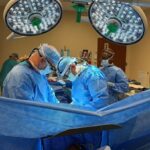
A major technological advancement in pulmonology is the introduction of the Ion endoluminal system, a robotic bronchoscopy machine that’s used to get biopsy samples of lung nodules.
Driven by two robotic arms, a 3.5mm catheter with a tiny camera is passed down an endotracheal tube. The pulmonologist uses two track balls on a control tower to guide the catheter down the airway. Once the target destination is reached, the catheter is parked to take a closer look and gather a tissue sample to be biopsied.
Holy Cross Health in Fort Lauderdale has a new bronchoscopy suite that was designed to accommodate this state-of-the-art system and recently purchased a fluoroscope which enables the physicians to do a 360-degree real-time scan of the patient while performing a biopsy using the Ion system.
The camera enables physicians to see precisely what is going on inside the body and adjust the needle’s position. The thinness of the catheter allows it to reach smaller nodules in areas of the lung that a traditional bronchoscopy procedure cannot.
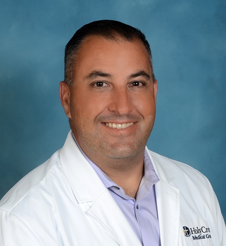
“It increases our percent yield,” Felix Hernandez, M.D., a pulmonologist at Holy Cross, explained. “It decreases our chances of missing a nodule because we’re literally able to directly see our biopsy tool exactly where it is in reference to that nodule, increasing our chances of getting a diagnostic sample. It’s a less invasive, higher-yield, less rate of complication way of figuring out what your lung nodule is.”
Holy Cross has used the Ion system on more than 80 patients. Fully FDA-approved and covered by insurance, Dr. Hernandez has yet to encounter a patient who chooses not to get a robotic bronchoscopy when presented with all options, which also includes a CT-guided biopsy performed by a radiologist or a more invasive wedge biopsy performed by a thoracic surgeon.
“Patients realize that a robotic bronchoscopy carries a lower rate of pneumothorax, or collapsed lung, compared to a CT-guided biopsy,” he said. “The other benefit of having a robotic bronchoscopy is that if the patient also has suspicious-looking enlarged lymph nodes in the chest, during the same procedure, we can just change scopes and also biopsy those lymph nodes.”
The robotic bronchoscopy can also determine what stage of cancer the patient has. A CT-guided biopsy only does a biopsy of the nodule, so a patient would have to return for a second procedure to get staged.
A person can be completely asymptomatic when a cancerous lung nodule is present. However, symptoms that should prompt an immediate visit to a pulmonologist include coughing up blood, pain when taking a deep breath in and unexpected weight loss.
Many patients don’t know they should be seen by a pulmonologist, so Dr. Hernandez and his team rely heavily on referrals from colleagues in other medical specialties such as primary care doctors, the emergency department, radiation oncologists, thoracic surgeons and more. At Holy Cross Health, they are educating these referring physicians about this emerging Ion technology because CT scans of the chest are often ordered by primary care physicians and emergency department doctors for a variety of reasons, including the increase in upper respiratory viruses.
If a lung nodule appears on the CT scan, “we want to optimize the care pathway for the patients,” Dr. Hernandez said. A pulmonologist should be the very next step to take a biopsy and determine if it is cancerous. An oncologist should not enter the picture until cancer is detected. If a patient is referred to an oncologist or thoracic surgeon first, they may undergo a more invasive procedure and/or delay a diagnosis because the oncologist would refer the patient to a pulmonologist before taking over the case.
The non-invasive, out-patient Ion endoluminal system robotic bronchoscopy procedure is done under anesthesia and can be completed in as little as 20 to 60 minutes.
To learn more or request an appointment, visit holycrossrobotics.com.