

Cleveland Clinic Florida uses the latest technological advancements in the fight against lung cancer
As technology continues to advance, patients are benefiting from breakthroughs, such as advanced robotic-assisted bronchoscopies, a new procedure that has revolutionized the way bronchoscopies are typically performed.
Cleveland Clinic Florida’s Weston Hospital and Indian River Hospital interventional pulmonologists are performing this procedure with precision, which allows them to accurately detect and diagnose lung cancer at an earlier stage. Pulmonologist Dr. Hermes Velasquez gives ample praise to this advancement.
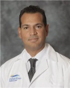
Dr. Hermes Velasquez
“First, it allows us to navigate more precisely. Secondly, we get more range, which allows us to reach further into the bronchial tube. And thirdly, it is more stable and once we reach our targeted place, we can anchor the device in that position, making it more advantageous to start the sampling process for biopsies,” said Dr. Velasquez.
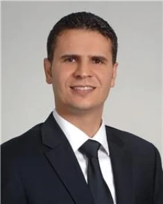
Dr. Ihab AlShelli
Similarly, Cleveland Clinic Weston interventional pulmonologist Dr. Ihab AlShelli explained, “With the robotic assisted bronchoscopy not only can we reach the area, but we can aim toward the exact location of the nodule and that will increase our chance to obtain a diagnosis,” said Dr. AlShelli. The technology greatly reduces the risk of false negatives, which means lung cancers can be caught and treated early, and patients are not undergoing repeated biopsies.
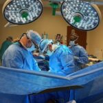
A robotically assisted bronchoscopy is performed on eligible patients diagnosed with a lung nodule or lung mass needing a biopsy to determine if the nodule is cancerous. This outpatient procedure based on electromagnetic navigation is performed under general anesthesia, and a CT scan is taken of the chest for the mapping tool on the day of the procedure. From there, the picture is recorded on a CD and placed into the robotic software which identifies any lesions in the CT scan to create a visual pathway for the surgeon to follow. Once the patient has been sedated, the flexible tube is placed in the endotracheal tube. The physician then uses a controller to manipulate a catheter and follows the outlined pathway to the nodule.
In a process called rapid on-site evaluation, an on-site pathologist looks at samples as they come in to determine whether they are compatible with cancer or other inflammatory conditions, explained Dr. Velasquez. If the pathologist only sees a healthy lung, the robot can guide the physician to another location to extract a better sample.
Patients with two or three lesions on the same side of the lung can be biopsied at the same time. Although samples cannot be taken from both sides of the lung for safety reasons, patients can return at a later date to continue the procedure, as there is no limit.
“Our goal is to detect lung cancer early in the game and ultimately save lives,” Dr. Velasquez said. “Lung cancer is curable if caught early, but unfortunately most lung cancer is discovered past the curable stage. That’s why it’s so important to get a CT lung cancer screening to make sure there are no lesions. By doing so, we can attack cancer in its earliest stages.”
To learn more about Cleveland Clinic Florida, visit my.clevelandclinic.org/florida.