We have reached an exciting turning point in the care of patients with breast cancer. We continue to see the commitment of multiple charitable organizations supporting breast cancer research and the innovations in technology come together to help us better deliver care, focused on the patient and not just the cancer. In short, there have been many advances across all aspects of breast cancer care, from diagnosis to treatment and survivorship.
Take the area of breast screening imaging, for example. Breast cancers are now being detected earlier than ever before, thanks to 3-D mammography technology that is helping radiologists find these tumors. Our radiologists are coupling 3-D mammography, also known as Tomosynthesis or Tomo, with C-view. This technology is FDA approved and provides patients with shorter scan time results in less compression time and with increased patient comfort. Other benefits include decreased recall rates for non-cancer cases, improved lesion margin visibility and precise localization of tumors.
From a breast surgical oncology perspective, the localization of tumors is extremely important. Most cancers cannot be felt by the surgeon, and in order to remove them, some form of localization is necessary. For the last 20 years, the gold standard to remove cancerous tumors has been a wire localization process in which a wire is placed in the breast the morning of the planned surgery by a radiologist. It is left protruding from the breast until the time of surgery, where it is removed along with the marked cancer. This process also makes for increased scheduling difficulty for the operating room because the radiologist and surgeon’s schedules need to be coordinated with precision. Ultimately, the process is time consuming for both the patient and surgeon.
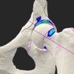
 Here at Memorial, we are now using SAVI Scout, a surgical guidance system that uses a small reflector, no larger than a staple, which is placed at the site of the cancer up to one week before surgery. A patient cannot tell that the reflector is in their breast. For the patient, this is more convenient and causes less anxiety. In the operating room, a special probe is used that sends out radar waves that hit the reflector and the reflector sends a signal back to the probe. When the probe gets the signal it emits a beep that lets the surgeon know exactly where both the reflector and the cancer are.
Other innovations that provide physical and psychological benefits to patients with breast cancer is having the option of having a nipple sparing mastectomy that preserves the nipple areola when a patient requires or desires a mastectomy. This procedure removes all of the breast tissue, but leaves all of the skin and nipple areola complex in place for use by the plastic surgeon for breast reconstruction.
Nipple sparing mastectomies used to be offered to women who met very strict criteria. Through many clinical trials, we learned that the risk of cancer recurrence is no higher than in women where the nipple is removed. Psychologically, it is a huge benefit for a woman to wake up from surgery, look in the mirror and still feel complete. As breast surgeons, we work closely with plastic surgeons to evaluate each patient to see if nipple sparing mastectomy could be an option.
Collaboration is key for us here at Memorial Cancer Institute. We have all the services for our breast cancer patients in our Memorial Breast Cancer Center. This type of collaboration between radiologists, oncologists, surgeons and many other members of the medical team allow us to provide one plan of care to each of our patients. That is patient-focused care.